Using artificial intelligence and a supercomputer, they are creating a 3D model of blood vessels that allows doctors to see the location of arteries and veins in the abdominal cavity before surgery. The development is a collaboration between experts from the Faculty of Electrical Engineering and Computer Science (FEECS), the IT4Innovations National Supercomputing Center at VSB – Technical University of Ostrava, and the University Hospital Ostrava as part of the LERCO project.
Each year, doctors at the Ostrava University Hospital perform around 340 surgeries on patients with colorectal cancer. In these procedures, knowing the exact location of blood vessels in the abdominal cavity is crucial. “The vascular system in the abdomen is highly variable and different in every individual. When a surgeon knows its course, they can plan the surgery better and reduce the risk of complications. Additionally, in colorectal cancer, it allows for an oncologically radical procedure, making the surgery as effective as possible for the patient,” explains Lubomír Martínek, Head of the Surgical Clinic at the University Hospital Ostrava.
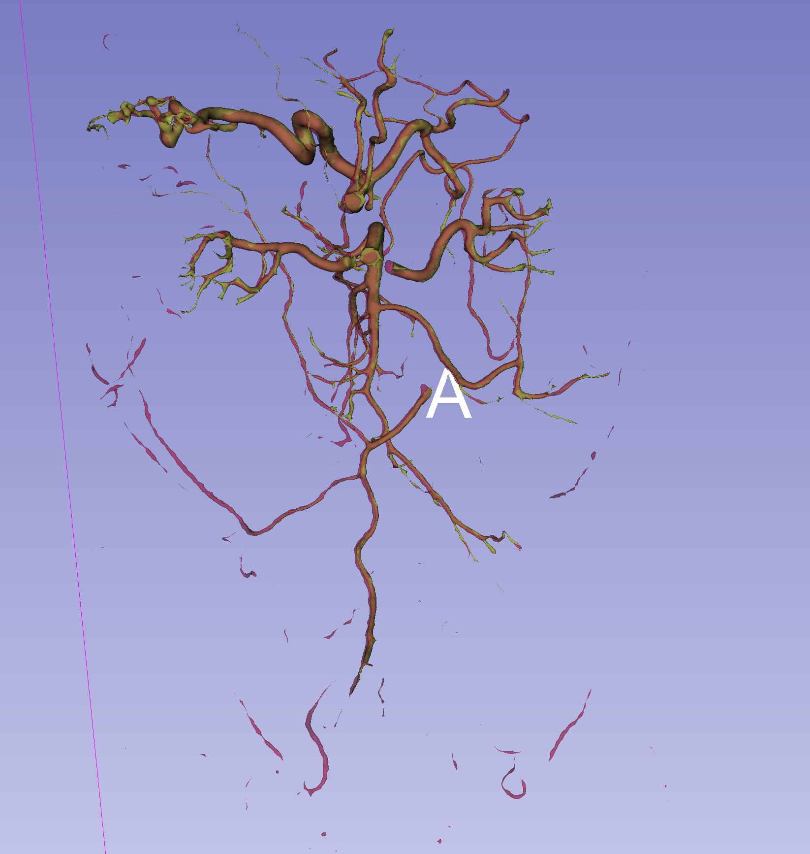
This is why researchers and doctors are working together to provide surgeons with a detailed 3D model of the blood vessels before surgery. “We selected 60 patients whose CT scans we annotated with blood vessels. It was very challenging because there are dozens of arteries and veins in the abdominal cavity. Manual segmentation — identifying and labeling all vessels larger than 2 mm — took 5 to 6 hours per patient,” says surgeon Jan Hrubovčák.
The result of the Ostrava doctors’ work is a unique dataset used by researchers from the Faculty of Electrical Engineering and Computer Science and IT4Innovations. “Our task was to develop a 3D segmentation algorithm based on deep learning. It learns to recognize vascular structures according to patterns defined by doctors,” explains Jan Kubíček from FEECS.
Training the algorithm is done on a supercomputer at IT4Innovations. “We are creating a neural network that can automatically identify the vascular system in the abdominal area from patient imaging data. While it takes a doctor several hours to segment the vessels, the current model can do it in 2.5 minutes for arteries, and the same time for veins,” adds Petr Strakoš from IT4Innovations.
The first results look very promising. A 3D model of arteries is already complete, and the team is now focusing on modeling veins and developing a specialized model for critical vessels located near tumors. The goal is to create a web application where a doctor can upload a specific patient’s data and receive an accurate 3D model of the vascular system within minutes.
This tool could bring many benefits for both doctors and patients. “More precise surgery planning, shorter operation time, and reduced risk of pre- and post-operative complications directly affect the prognosis and chance of recovery for the patient. If the algorithm could segment vessels in real time, it could find wide application not only in research but also in routine clinical practice,” adds Chief Physician Martínek.